
Клинические и радиологические предикторы развития эпилепсии у пациентов с цереброваскулярной патологией: обзор литературы с систематическим анализом данных
Клинические и радиологические предикторы развития эпилепсии у пациентов с цереброваскулярной патологией: обзор литературы с систематическим анализом данных

Аннотация
Введение. Цереброваскулярные заболевания являются одними из ведущих причин приобретенной эпилепсии у взрослых и пожилых пациентов. Несмотря на достижения в лечении острого инсульта и нейровизуализации, надежное прогнозирование развития эпилепсии после цереброваскулярных событий остается сложной задачей из-за многофакторного характера эпилептогенеза после инсульта.
Цель. Обобщить и критически проанализировать имеющиеся данные о клинических и нейровизуализационных предикторах развития эпилепсии после цереброваскулярных заболеваний.
Методы. Был проведен нарративный обзор с систематическим поиском литературы в базах данных PubMed/MEDLINE, Scopus, Web of Science и ScienceDirect. Были проанализированы публикации с 2000 по 2025 год, с особым акцентом на исследования за последние 10–15 лет. Были оценены клинические факторы риска, нейровизуализационные характеристики (КТ и МРТ) и электроэнцефалографические данные, связанные с эпилепсией после инсульта.
Результаты. Наиболее часто упоминаемыми клиническими предикторами эпилепсии после инсульта были ранние симптоматические судороги, тяжесть инсульта и геморрагический подтип инсульта. К нейровизуализационным признакам с наибольшей прогностической ценностью относились кортикальное поражение, большой объем очага поражения, геморрагическая трансформация, кортикальный сидероз и хронические структурные изменения, такие как глиоз и кортикальная атрофия. Аномалии ЭЭГ, особенно эпилептиформные разряды и очаговое замедление, предоставляли дополнительную прогностическую информацию в сочетании с клиническими и радиологическими данными. Существующие клинические шкалы оценки риска продемонстрировали умеренную прогностическую эффективность и ограниченную обобщаемость.
Выводы. Эпилепсия после инсульта является результатом сложного взаимодействия между клинической тяжестью, структурным повреждением головного мозга и вторичной реорганизацией нейронных сетей. Мультимодальный подход, интегрирующий клинические данные, передовые методы нейровизуализации и электрофизиологические данные, может улучшить индивидуальную стратификацию риска и раннее выявление пациентов с высоким риском развития эпилепсии.
1. Введение
Cerebrovascular diseases (CVD) remain a leading cause of disability and mortality worldwide and a significant factor in the development of symptomatic epilepsy in adults and elderly patients. According to epidemiological studies, up to 30–40% of cases of newly diagnosed epilepsy in the elderly are associated with cerebrovascular lesions, primarily ischemic stroke and intracerebral hemorrhage
, . The development of epilepsy against a background of cerebrovascular pathology significantly worsens functional outcome, increases the risk of rehospitalization, and reduces the quality of life of patients.Post-vascular epilepsy is a heterogeneous and multifactorial condition that develops as a result of the interaction between clinical characteristics, structural brain damage, and neurophysiological changes , . Despite substantial advances in neuroimaging technologies and modern stroke management, reliable criteria allowing accurate prediction of epilepsy development in individual patients remain insufficiently established. This limitation complicates early risk stratification and delays identification of patients requiring close neurological monitoring.
Among the most consistently reported predictors of epilepsy following cerebrovascular events are stroke type, cortical localization of the lesion, lesion volume, presence of hemorrhagic components, early symptomatic seizures, and severity of chronic structural brain alterations . Modern MRI techniques, including FLAIR, susceptibility-weighted imaging (SWI), and diffusion-weighted imaging (DWI), allow detection not only of acute ischemic or hemorrhagic changes but also of subclinical markers of secondary epileptogenesis, such as cortical gliosis, cerebral microbleeds, cortical siderosis, and disturbances of structural connectivity , , . Therefore, systematic evaluation of clinical and radiological predictors of epilepsy in patients with cerebrovascular disease remains highly relevant.
Objective. To analyze and summarize current data on clinical and neuroimaging predictors of epilepsy development in patients with cerebrovascular disease.
2. Materials and Methods
This study was conducted as a narrative review with a structured literature search focusing on clinical and radiological predictors of epilepsy following cerebrovascular disease.
A comprehensive search of international electronic databases, including PubMed/MEDLINE, Scopus, Web of Science, and ScienceDirect, was performed. Publications from January 2000 through December 2025 were included, with particular emphasis on studies published during the last 10–15 years to reflect contemporary diagnostic approaches and advances in neuroimaging.
The following keywords and their combinations were used: post-stroke epilepsy, vascular epilepsy, cerebrovascular disease, clinical predictors, neuroimaging predictors, MRI biomarkers, hemorrhagic transformation, early seizures, electroencephalography, machine learning, artificial intelligence, and radiomics. Additionally, reference lists of key review articles and original studies were manually screened to identify relevant publications not retrieved during the primary search.
The review included:
· observational cohort studies (retrospective and prospective);
· systematic reviews and meta-analyses;
· studies involving adult patients (≥18 years);
· investigations focusing on epilepsy associated with ischemic stroke,
· intracerebral hemorrhage, or chronic cerebrovascular disease;
· studies analyzing clinical predictors, neuroimaging findings, and electroencephalographic characteristics related to epilepsy development.
Exclusion criteria were:
· case reports and small case series (<10 patients);
· studies limited to pediatric populations;
· investigations addressing seizures caused exclusively by metabolic or toxic disturbances;
· studies lacking clear differentiation between early symptomatic seizures and late post-stroke epilepsy;
· articles without adequate methodological description or unavailable full text.
Data Extraction and Synthesis
From the included studies, data were extracted and qualitatively synthesized regarding study design, sample size, type of cerebrovascular pathology, timing of seizure occurrence, clinical predictors (age, stroke severity, neurological status, early seizures), neuroimaging characteristics (lesion location, size, cortical involvement, hemorrhagic components, chronic structural changes), electroencephalographic findings when available, and statistical or machine learning approaches used for epilepsy risk prediction.
Predictors were categorized into clinical, radiological, and combined multimodal groups. Factors associated with early symptomatic seizures were analyzed separately from those related to late post-stroke epilepsy. Due to heterogeneity in study design, outcome definitions, and reporting methods, quantitative meta-analysis was not performed.
Additional clinical risk factors included a more severe course of stroke, pronounced neurological deficit in the acute period, impaired consciousness, and a relatively younger age of patients, which is especially characteristic of the development of late post-vascular epilepsy (Fig. 2).
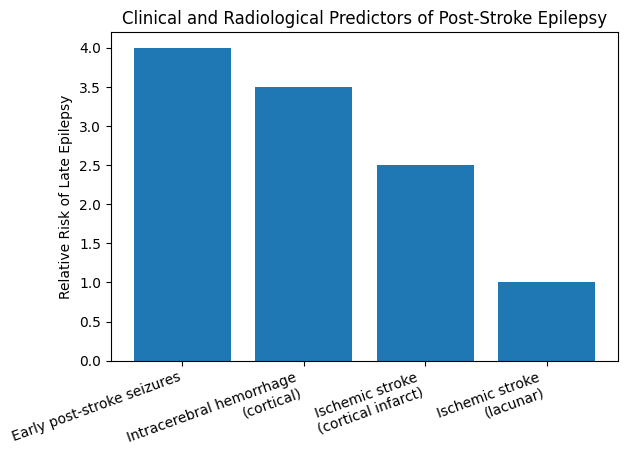
Risk of epilepsy development according to the type of cerebrovascular event
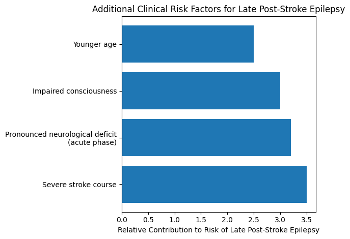
Clinical predictors associated with an increased risk of post-stroke epilepsy
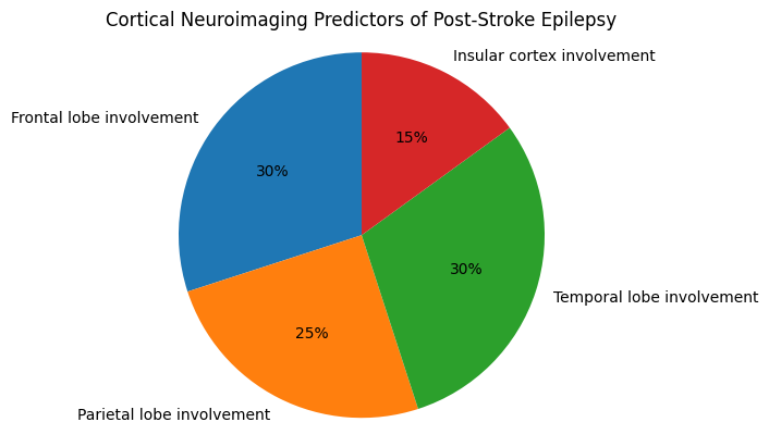
Neuroimaging predictors of post-stroke epilepsy
3. Main results
As shown in figure 3, neuroimaging characteristics of the lesion focus were among the most reproducible predictors of epilepsy development. All analyzed studies emphasized the key role of the cortical location of the stroke. Damage to the frontal, parietal, and temporal lobes, as well as involvement of the insular cortex, significantly increased the risk of developing an epileptogenic focus: an acute injury → structural damage → network reorganization → epileptogenesis.
Lesion volume also had prognostic significance: large infarcts and extensive intracerebral hemorrhages were associated with a higher incidence of late-onset epileptic seizures. The presence of a hemorrhagic component, including hemorrhagic transformation of ischemic stroke, was an independent risk factor, particularly with the detection of cortical siderosis and microbleeds in SWI modes. Chronic structural changes in brain tissue, such as post-stroke gliosis, cortical atrophy, and pronounced leukoaraiosis, also contributed to the development of epileptic activity, reflecting secondary epileptogenesis and neural network disruption. Several studies have emphasized the additional prognostic value of electroencephalography. The detection of epileptiform activity, periodic discharges, or pronounced focal slowing in the area of vascular lesion correlated with an increased risk of developing late-onset epilepsy. However, the sensitivity of EEG in the acute period of stroke remained limited, which emphasizes the need for its interpretation in combination with clinical and radiological data (Table 1).
Multimodal predictors of late post-stroke epilepsy based on structural neuroimaging and electrophysiological findings
Domain | Predictive Factors | Imaging / Diagnostic Modality | Prognostic Significance |
Lesion volume and type | Large ischemic infarcts | CT / MRI | Higher incidence of late epileptic seizures |
Extensive intracerebral hemorrhage | CT / MRI | Increased risk of post-stroke epilepsy | |
Hemorrhagic components | Hemorrhagic transformation of ischemic stroke | CT / MRI | Independent risk factor for late epilepsy |
Cortical siderosis | MRI (SWI) | Strong association with epileptogenesis | |
Cerebral microbleeds | MRI (SWI) | Marker of increased seizure susceptibility | |
Chronic structural brain changes | Post-stroke gliosis | MRI (T2/FLAIR) | Substrate for secondary epileptogenesis |
Cortical atrophy | MRI (volumetric / visual assessment) | Network disruption and seizure generation | |
Electrophysiological findings | Severe leukoaraiosis | MRI (FLAIR) | Impaired connectivity and increased epileptic risk |
Epileptiform discharges | EEG | Strong predictor of late epilepsy | |
Periodic lateralized discharges | EEG | Associated with higher seizure recurrence | |
Focal slowing in lesion area | EEG | Reflects cortical dysfunction and epileptogenicity | |
Integrated assessment | Combined clinical, radiological and EEG markers | Multimodal approach | Improved risk stratification and prediction accuracy |
4. Discussion
The findings of this narrative review indicate that epilepsy developing after cerebrovascular disease is the result of a multifaceted interaction between the severity of acute vascular injury, structural brain alterations, and secondary processes of network reorganization. Across the analyzed literature, the most stable and reproducible predictors included cortical involvement, the presence of hemorrhagic components within the lesion, and the occurrence of early symptomatic seizures, which is consistent with contemporary concepts of post-stroke epileptogenesis , , .
Cortical localization of cerebrovascular damage was identified as the most powerful predictor of epilepsy development. The cerebral cortex serves as the principal substrate for epileptic activity, and vascular injury to cortical structures initiates a cascade of pathological changes including neuronal loss, synaptic remodeling, disruption of excitatory–inhibitory balance, and activation of inflammatory signaling pathways , . Lesions affecting frontal, temporal, parietal, and insular regions were consistently associated with elevated epilepsy risk, whereas purely subcortical or lacunar infarctions demonstrated substantially lower epileptogenic potential , .
The presence of hemorrhagic components, such as primary intracerebral hemorrhage or hemorrhagic transformation of ischemic infarction, was another significant predictor of late epilepsy , . Pathophysiological mechanisms include iron accumulation, persistent cortical irritation caused by blood breakdown products, and disturbances in ionic homeostasis. Neuroimaging markers including cortical siderosis and cerebral microbleeds, particularly detected using susceptibility-weighted imaging, reflect these processes and have been repeatedly associated with increased seizure susceptibility , .
Early symptomatic seizures occurring during the acute phase of stroke were among the strongest predictors of later epilepsy development , . These seizures likely represent not only an acute reaction to structural injury but also an indicator of already initiated epileptogenic mechanisms within vulnerable neuronal networks. Their presence suggests irreversible alterations in cortical excitability that predispose to recurrent unprovoked seizures.
Chronic structural changes also play an essential role in epileptogenesis. Post-stroke gliosis, progressive cortical atrophy, and severe leukoaraiosis have been consistently linked to increased epilepsy risk , . These changes reflect long-term disruption of neural connectivity, maladaptive neuroplasticity, and a gradual reduction of seizure threshold, which explains the delayed onset of epilepsy months or years after the primary vascular event.
Electroencephalography provides additional prognostic information when interpreted together with clinical and imaging data. Epileptiform discharges, periodic lateralized discharges, and pronounced focal slowing in regions corresponding to vascular lesions have been associated with an increased probability of epilepsy development , . However, EEG sensitivity during the acute stroke period remains limited, emphasizing the need for multimodal assessment combining neurophysiological and structural data.
Existing clinical prediction models, such as the SeLECT and CAVE scores, represent important steps toward standardized risk estimation of post-stroke epilepsy , . Nevertheless, their predictive accuracy remains moderate, and they do not fully account for the complexity of structural and electrophysiological factors contributing to epileptogenesis. In particular, these models insufficiently incorporate chronic brain changes and advanced neuroimaging biomarkers.
Traditional statistical approaches have significantly contributed to identifying epilepsy predictors after stroke but remain limited in capturing the heterogeneity of cerebrovascular lesions and individual disease trajectories , . This highlights the need for integrated clinical evaluation combining neurological examination, imaging findings, and electrophysiological assessment in routine practice.
Clinical Implications
From a clinical standpoint, early recognition of patients at high risk for epilepsy following cerebrovascular disease is of considerable importance. Individuals with cortical involvement, hemorrhagic components, and early symptomatic seizures require closer neurological follow-up, repeated EEG evaluation, and careful monitoring for seizure recurrence. Comprehensive interpretation of MRI findings, including markers of chronic brain injury, may further enhance individualized risk stratification and improve patient counseling strategies , .
Limitations
Several limitations of this review should be acknowledged. First, substantial heterogeneity in study designs, outcome definitions, and follow-up durations prevented the performance of a quantitative meta-analysis. Second, most included studies were observational, which restricts causal inference. Third, differences in neuroimaging protocols and EEG timing across studies may have influenced the reported predictive value of certain markers. Finally, publication bias cannot be excluded, as studies reporting significant associations are more likely to be published.
5. Conclusion
Epilepsy developing after cerebrovascular disease arises from a complex interplay between acute vascular injury, chronic structural brain alterations, and progressive network reorganization processes , . The most consistent predictors identified across studies include cortical localization of lesions, the presence of hemorrhagic components, and early symptomatic seizures , .
Early identification of high-risk patients using an integrated approach that combines clinical assessment, neuroimaging findings, and electrophysiological data may improve monitoring strategies, support preventive interventions, and facilitate individualized patient counseling , .
Disclosure Statement
During the preparation of this work, the authors used ChatGPT to assist in identifying relevant scientific publications. Following the use of this tool, all content was carefully reviewed, revised, and verified by the authors, who assume full responsibility for the final manuscript.